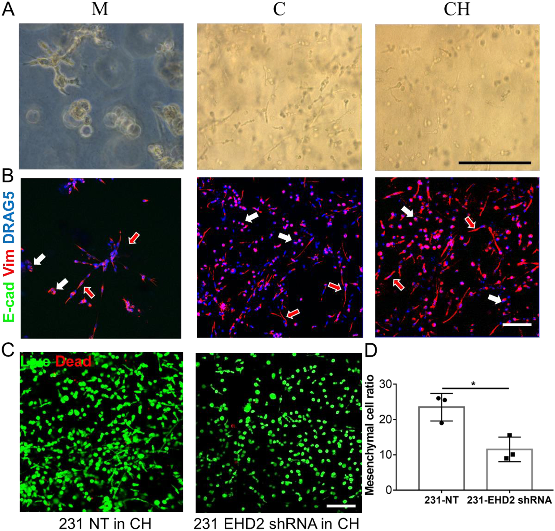
Figure 6.

Evaluation of MDA-MB-231 human breast cancer cells behavior and phenotype in embedded bioprinted models of three different bioinks. A) MDA-MB-231 showed a mixture of non-invasive epithelial and mesenchymal phenotypes in all three bioinks at day 7. B) IF staining of E-cadherin (not detected) and vimentin proteins on MDA-MB-231 cells in three bioinks. The cells or cell clusters with epithelial phenotypes were indicated by the all-white arrow, while the red-in-white arrow represented the mesenchymal phenotypes. C) Live and dead staining of MDA-MB-231 cells transfected with NT and EHD2 shRNA in CH model. D) Quantification of mesenchymal phenotype ratio of two modified MDA-MB-231 cells in the bioprinted CH model (n=3, *p<0.05). Scale bar: black, 250 μm, white, 100 μm
