
Figure 2. Lower resolution histopathologic features of mouse lungs 8 days following infection with SARS-CoV (strain Urbani).
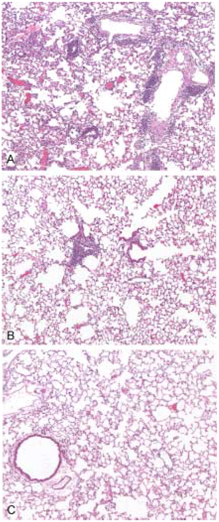
The lungs of mice receiving normal mouse serum show multifocal and extensive perivascular and interstitial inflammatory cell infiltrates (A). In contrast, mice receiving a 1:4 dilution of hyperimmune SARS-CoV antiserum show only occasional small foci of perivascular infiltrates (B) and the mice that received undiluted hyperimmune antiserum showed no significant pulmonary inflammation (C). Hematoxylin and eosin stain. Original magnifications × 25.