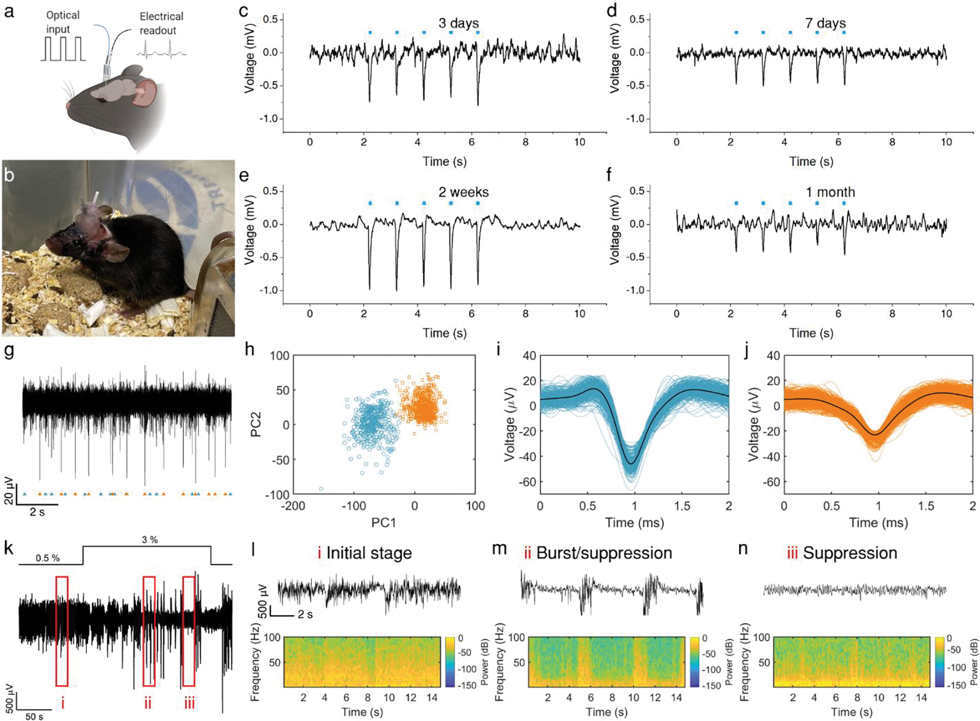
Figure. 3. Simultaneous optoacoustic stimulation and electrophysiological recording by implanted mFOE in mouse hippocampus.

a. Illustration of the mFOE enabled bidirectional neural communication using laser signal as input and electrical signal as readout. b. mFOE was implanted into hippocampus of a wild type C57BL/6J mouse. c-f. Simultaneous optoacoustic stimulation and electrophysiological recording performed at 3 days (c), 7 days (d), two weeks (e) and one month (f) after implantation. Blue dots the laser pulse trains. For each laser train: 50 ms burst of pulses, pulse energy of 41.8 μJ, laser repetition rate 1.7 kHz. g. Part of the filtered spontaneous activity containing two separable group of spikes recorded by mFOE electrode at one month after implantation. h. Principal-components analysis (PCA) of the two group of spikes. i-j. Waveform of two group of spikes in h. k. Local field potential (LFP) recorded by mFOE one month after implantation with an alternating anaesthesia level (0.5–3% v/v isoflurane). l-n. different LFP responses induced by varying the concentration of isoflurane: l corresponds to the initial stage (0.5% of isoflurane level); m corresponds to the burst/suppression transition stage (after increasing the isoflurane level to 3%); n corresponds to the suppression stage (the isoflurane level was maintained at 3% and took effect).
