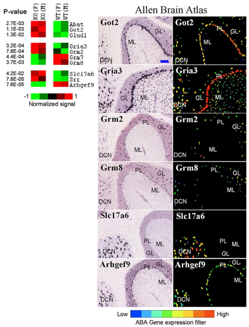
Figure 1.

Genes differentially expressed in the cerebellum and involved in GABA and glutamate neurotransmission. Left, Differential expression between knock-out (KO) and wild-type (WT) lines is shown in pseudocolor with associated p values from a t test. Average values for Pittsburgh (P) and Merck (M) lines are shown. Right, ABA images show regional distribution of six selected transcripts. ML, Molecular layer; PL, Purkinje cell layer; GL, granule layer; DCN, deep cerebellar nuclei. Got2, Gria3, and Abat (not shown) are expressed in all four subregions examined. Grm2 is expressed in Golgi cells in the GL. Grm7 is predominantly expressed in Purkinje neurons, whereas Grm8 is detected in ML, DCN, and Golgi cells (Berthele et al., 1999). VGLUT2 (Slc17a6 ) was detected only in the DCN, and collybistin (Arhgef9) was present in PL, GL, and DCN. Glud1 and Srr are preferentially expressed in astrocytes (Zaganas et al., 2001; Ribeiro et al., 2002). The blue bar in the top left image represents ~100 μm. All images are copied and modified with permission from Allen Institute for Brain Science.
