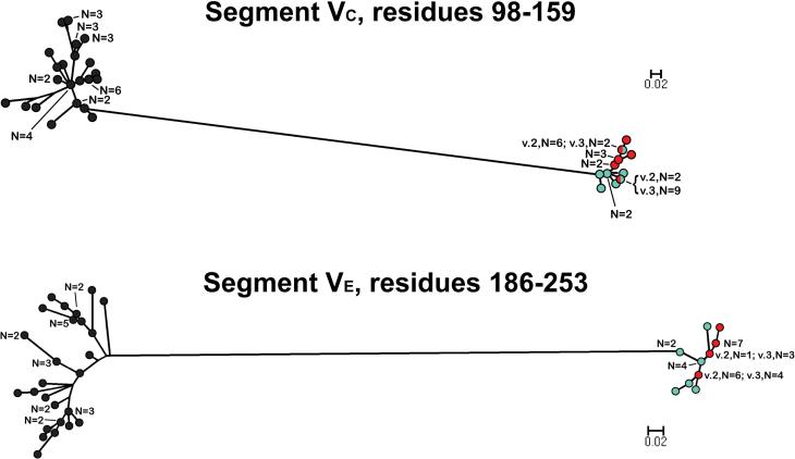
Figure 4.
Phylograms of unique fHbp amino acid sequences in variable segments VC (residues 98-159) and VE (residues 186-253). The colors of the circles in for segments in each variant group correspond to those described in the legend of Figure 3. E.β.10 refers to peptide ID no. 82 with an exceptional junctional point (See text). The scale bars indicate 2 amino acid changes per 100 residues.

