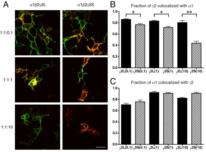
Figure 2.
γ2S subunits can also express independently, even in the presence of α1 and β2 subunits. A. Selected images from colocalization experiments performed on HEK293 cells transfected with α1β2γ2 cDNAs at 1:1:0.1, 1:1:1 and 1:1:10 ratios. Cells were labeled with 565 nm quantum dot-labeled antibodies (green) for α1 subunits and 655 nm (far red, pseudocolored red) for γ2 subunits (see Materials and Methods). Colocalized signal ranges from yellow-green to orange-red. Bar = 20 μm. B. Measurements of colocalization of γ2L or γ2S with α1 signal. At each transfection ratio, γ2S subunits were significantly different from γ2L subunits in the amount of red signal that was not colocalized with green (α1) signal. Data are mean ± s.e.m. for n ≥ 50 cells per condition, * p < 0.01, ** p < 0.001. C. Control measurements of α1 colocalization with γ2L or γ2S. At no transfection ratio does γ2S differ significantly from γ2L for this measurement.

