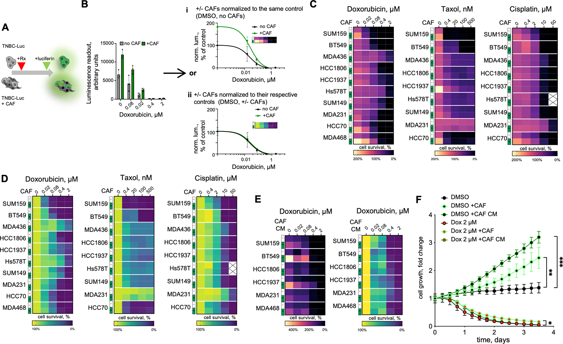
Figure 1. CAFs facilitate TNBC proliferation in vitro.

A. Experiment diagram for the chemosensitivity sensitivity assay. Luciferase-labeled TNBC cells are cultured in the presence or absence of unlabeled CAFs in the presence of doxorubicin or DMSO vehicle control. Only TNBC cells directly contribute to the viability signal. B. Normalization schemata for the data analyses. Raw data from the viability assay can be normalized to either i) the DMSO control signal of cells cultured without CAFs, or ii) with separate normalization of the control and CAF co-cultures to their respective DMSO controls. C, D. Heatmap summaries of the impact of CAF co-cultures on the sensitivity of the indicated chemotherapeutic agent in a panel of TNBC cell lines, normalized as i) or ii) in panel B, respectively. E. Heatmap summaries of the impact of CAF CM on doxorubicin sensitivities of the indicated TNBC cell lines. F. Impact of CAFs and CAF CM on the growth of GFP-labelled MDA468 cells following 24 hours of doxorubicin exposure, measured by time-lapse microscopy. Statistical analyses of indicated differences were performed with a paired 2-tail t-test, comparing confluency value at each of the time point *** p=0.0007, ** p=0.003, * p=0.0102.
