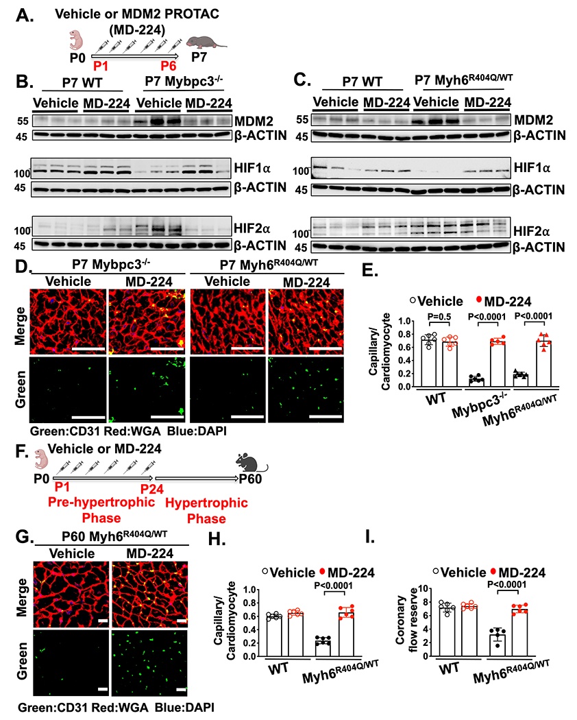
Figure 7. Chemical inhibition of MDM2 prevents microvascular dysfunction in two distinct HCM models.

(A) Schematic of injections of vehicle or MDM2 PROTAC (MD-224) from P1 to P6. (B) Immunoblots for MDM2, HIF1α and HIF2α in LV tissue from P7 WT or Mybpc3−/− injected with vehicle or MD-224. (C) Immunoblots for MDM2, HIF1α and HIF2α in LV tissue from P7 WT and Myh6R404Q/WT injected with vehicle or MD-224. (D) Representative immunohistochemistry images for CD31 (green) co-stained with WGA (red) in LV tissue from P7 Mybpc3−/− and Myh6R404Q/WT injected with vehicle or MD-224 from P1-P6. Nuclei are blue (DAPI). Scale bars=50 μm. (E) Capillary to cardiomyocyte ratios in LV tissue from P7 WT vehicle (n=6), WT MD-224 (n=6), Mybpc3−/− vehicle (n=6), Mybpc3−/− MD-224 (n=5), Myh6R404Q/WT vehicle (n=7) and Myh6R404Q/WT MD-224 (n=6) mice. All groups injected from P1-P6 with either vehicle or MD-224. Minimum 200 cardiomyocytes/sample. (F) Schematic of WT or Myh6R404Q/WT mice injected with vehicle or MDM2 PROTAC (MD-224) from P1 to P24 and then analyzed at P60. (G) Representative immunohistochemistry images for CD31 (green) co-stained with WGA (red) in LV tissue from P60 Myh6R404Q/WT mice injected with vehicle or MD-224 from P1 to P24. Nuclei are blue (DAPI). Scale bars=15 μm. (H) Capillary to cardiomyocyte ratios in LV tissue from P60 WT vehicle (n=6), WT MD-224 (n=6), Myh6R404Q/WT vehicle (n=6) and Myh6R404Q/WT MD-224 (n=6) mice. All groups injected from P1 to P24 with vehicle or MD-224. Minimum 140 cardiomyocytes/sample. (I) Coronary flow reserve was calculated in P60 WT vehicle (n=6), WT MD-224 (n=6), Myh6R404Q/WT vehicle (n=5), and Myh6R404Q/WT MD-224 (n=6) mice. All results are shown as mean±SEM. Student’s t-test or Welch’s t-test were utilized for E, H, and I.
