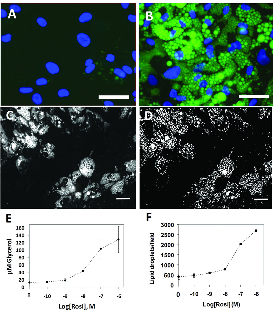
Figure 1. Development of the Lipid Droplet algorithm.
Human primary preadipoctes were cultured in 96-well dishes in the absence or presence of 1 µM rosiglitazone for 14 days; the cells were then fixed, permeabilized and labeled for nuclei and lipid droplets using Vala’s Lipid Droplet reagent kit, then imaged using a Beckman IC 100 Image Cytometer outfitted with a 40X objective. A, Control cells cultured in the absence of rosiglitazone; nuclei are blue and lipid droplets are green. B, Cells exposed to 1 µM rosiglitazone. C, Gray scale image of the lipid droplet image obtained from rosi-treated cells. D, A binary mask is shown, derived from C, in which the individual lipid droplets are identified. E, The effect of rosi on triglycerol content is shown (each symbol is the mean ± SD, n=8 wells). F, The effect of rosi on lipid droplets/field of view is shown, as quantified by the Lipid Droplet algorithm (each symbol is the Mean ± range, n= 2 wells). Scale bars are 50 µm.

