
Figure 2:
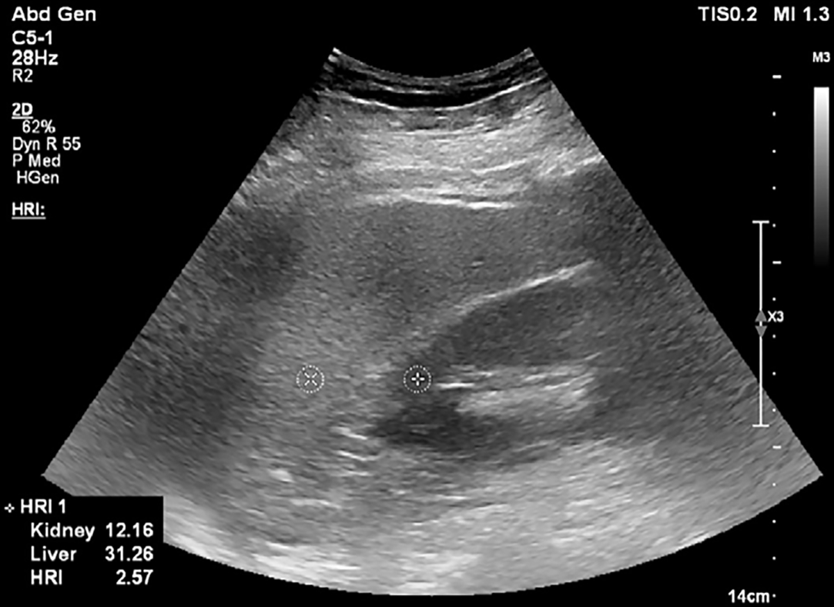
B-mode US image at the level of the pouch of Morison in a 59-year-old female patient with nonalcoholic steatohepatitis and grade 2 hepatic steatosis. Two circular regions of interest are placed on the kidney cortex and liver tissue at the same depth. Hepatorenal index (HRI) can be calculated on US systems with HRI quantification software, or images can be exported in Digital Imaging and Communications in Medicine, or DICOM, format and region of interest circles can be drawn with a DICOM viewer. In this image, the local region of interest pixel brightness values and the HRI value are presented in the bottom left corner. Reproduced, with permission, from the Non-Invasive Biomarkers of Metabolic Liver Disease, or NIMBLE 1.1, study (18).