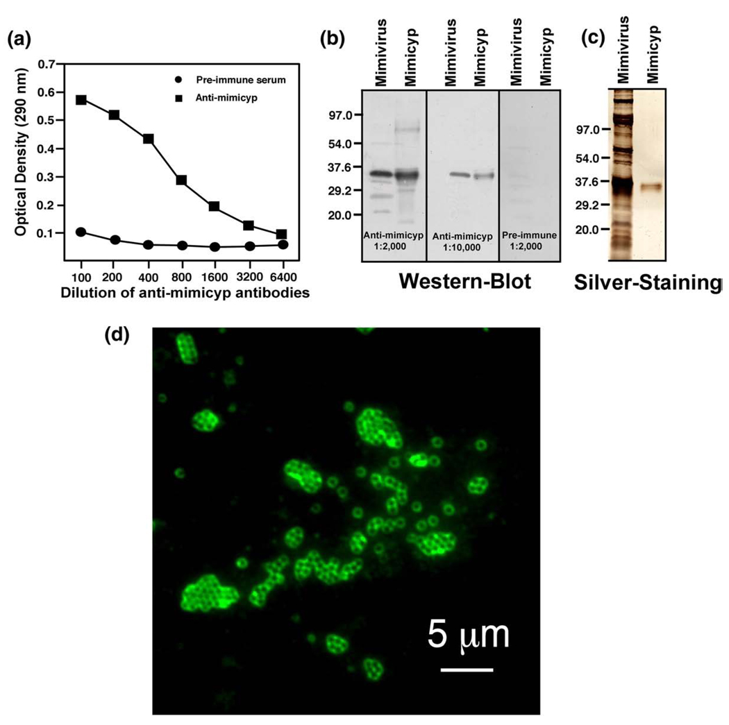
Figure 5.
Mimicyp expression and location on the mature Mimivirus virion. (a) ELISA detection of mimicyp in Mimivirus extracts using the anti-mimicyp polyclonal antibodies. (b) As indicated by immunoblots using anti-mimicyp antibodies (1:2000 and 1:10,000 antibody dilutions), mimicyp is found in viral extracts (indicated as Mimivirus) and migrates similarly to recombinantly expressed mimicyp (indicated as Mimicyp). As a control, pre-immune serum was also probed and exhibited an absence of any detectable mimicyp. (c) Those proteins recognized by anti-mimicyp other than the recombinant protein itself represent a minority, since they are not detected upon silver staining on SDS–PAGE. (d) Mimicyp is located on the outside of mature Mimivirus virions, as detected by immunofluorescent staining of viral particles with anti-mimicyp polyclonal antibodies.

