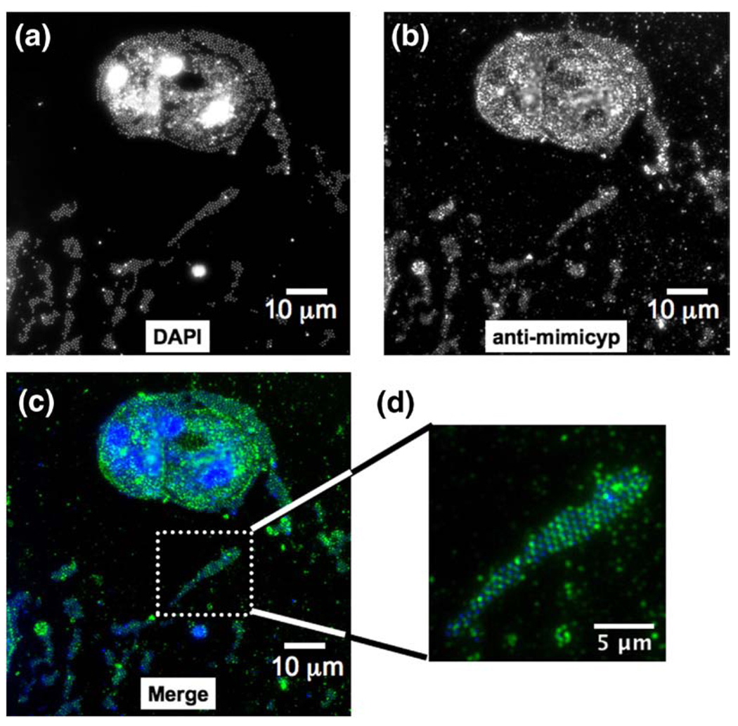
Figure 6.
Mimicyp expression during the Mimivirus infection of A. polyphaga. (a) DAPI staining allows identification of viral particles. Large fluorescent areas have recently been identified as viral factories within the infected hosts.46 (b) Anti-mimicyp polyclonal antibody staining shows that mimicyp is highly expressed within the infected amoebal hosts as well as enveloping released virions. (c) Color coded merged Figure showing DAPI staining (blue), anti-mimicyp antibody staining (green), and merged (light blue). (d) Expanded view of released virions. Staining proceeded as described 16 h post infection46 and no fluorescence was observed in the control using preimmune serum.

