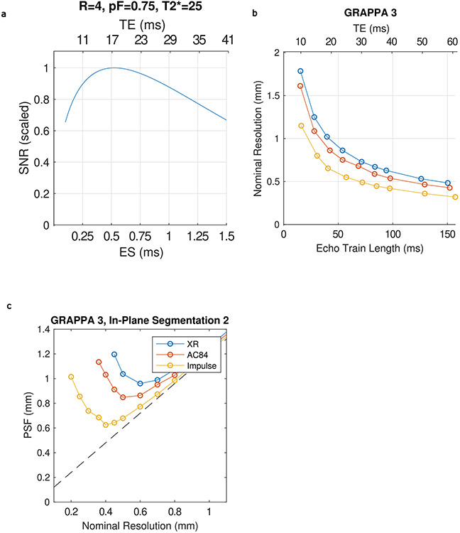
Extended Data Fig. 5 ∣. Gradient coil performance and EPI.
(a), Simulation of the effects of Echo Time (TE) and echo spacing on SNR in EPI. (b), achievable nominal resolution for different minimum TEs for three different gradient coil performances when using GRAPPA acceleration of 4. (c), Point Spread Function (PSF) on image phase encoded axis (FWHM of Fourier transform of the modulation transfer function associated with T2* decay across the image readout, modeled using a T2* of 25 ms) versus nominal resolution for 3 different gradient coil performances for GRAPPA 3 combined with In-plane Segmentation (for an equivalent acceleration of 6). Dashed black line shows minimum achievable PSF for a given nominal resolution assuming no T2* blurring. (d) Comparison of SNR for central (blue) and peripheral (red) regions of the brain. Gmax, SR (mT/m, T/m/s) XR and SC72 whole-body gradient (80,200), AC84 head only gradient (80,400), Impulse head-only gradient (200, 900).

