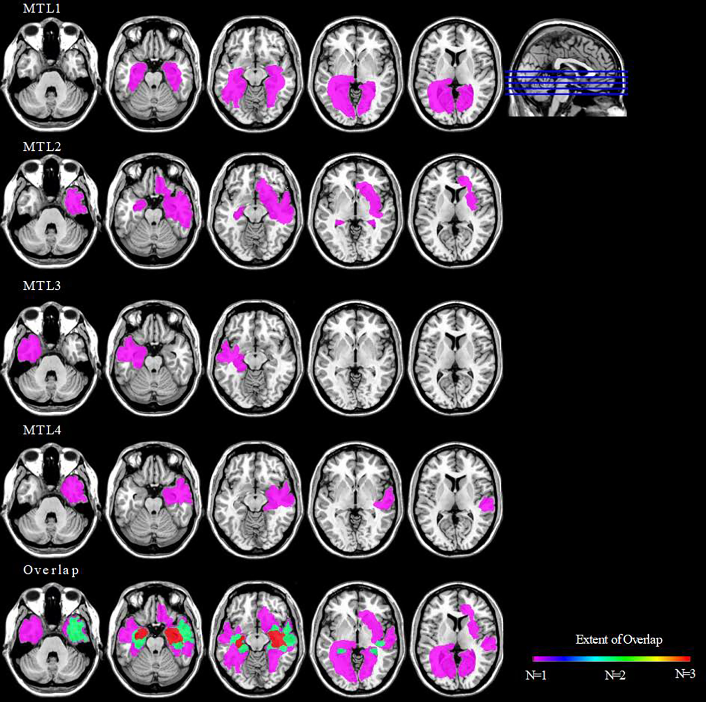
Figure 1. Lesion location and overlap among the individuals with medial temporal lobe amnesia.

As shown here, individuals had damage to bilateral (MTL1 and MTL2) or unilateral (MTL3 and MTL4) hippocampal and surrounding medial temporal lobe structures. Lesions extended into the cortex in all cases, although there was limited overlap. Lesions were manually drawn on the ch2 atlas in MRICron and are shown such that left reflects the left side of the brain. The color bar shows the amount of overlap in lesion location across the four individuals.
