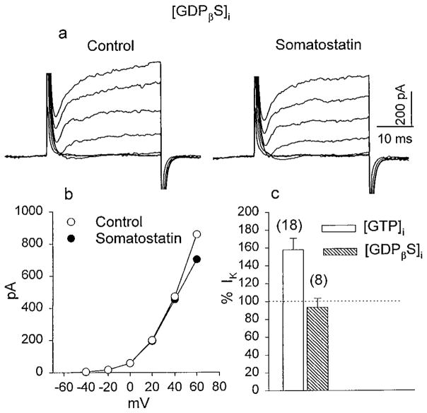
Figure 4.
The effect of GDPβS on SRIF-induced changes in IK. a, K+ currents were recorded with a patch pipette containing 500 μM GDPβS in control solution and after a 5 min exposure to 0.5 μM SRIF. b, The I–V relationship of IK obtained from the experiments described in a indicates that SRIF failed to alter IK in the presence of GDPβS. c, The histogram summarizes SRIF-induced changes in IK when the internal solution contained GTP (open bar) and when GTP was replaced by GDPβS (hatched bar) to block G-protein activation.

