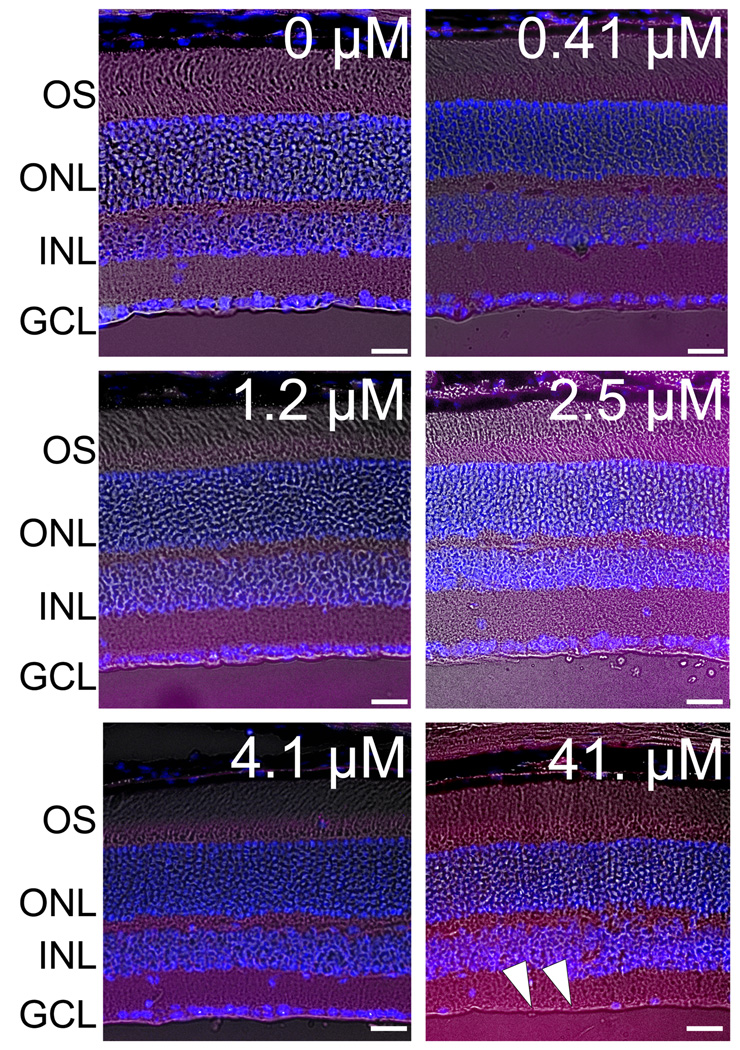
Figure 4. Histopathological examination of radial sections of mice eyes 7 weeks after intravitreal injection of balanced salt solution (BSS) or incrementing concentrations of caspofungin in BSS.
Superimposed on a phase contrast image are DAPI stained nuclei in various cell layers (blue) and eosin stained cell membrane (red) prominently seen in the plexiform layers. No retinal abnormalities were noted in the retinae injected with BSS alone or 0.41 µM to 4.1 µM of caspofungin dissolved in BSS. Eyes injected with 41. µM of caspofungin showed loss of cells in the ganglion cell layer (arrowheads). OS, outer and inner segments of the photoreceptors; ONL, outer nuclear layer; INL, inner nuclear layer; and GCL, ganglion cell layer. Scale bar = 20 µm.

