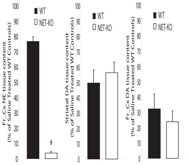
Figure 1. Treatment with αMT induces cortical NE depletion in NET-KO mice.
Treatment with αMT (250 mg/kg) reduced cortical NE content in NET-KO mice to less than 5% of that observed in saline treated WT control mice (left); n= 12 and 11 for saline treated WT and NET-KO, respectively, and n= 6 and 5 for αMT treated WT and NET-KO, respectively. Treatment with αMT (250 mg/kg) produced an equivalent 40–50% reduction in striatal dopamine content in WT and NET-KO mice (right); n= 12 and 10 for saline treated WT and NET-KO, respectively, and n= 4 and 4 for αMT treated WT and NET-KO, respectively. Treatment with αMT (250 mg/kg) produced an equivalent 70–75% reduction in cortical dopamine content in WT and NET-KO mice (right); n= 6 and 5 for saline treated WT and NET-KO, respectively, and n= 5 and 5 for αMT treated WT and NET-KO, respectively. Importantly, there was no difference cortical dopamine levels observed between saline treated NET-KO mice and WT controls; p>0.05. Catecholamine levels were measured 2 hours after treatment with saline or αMT as previously described (Wang et al., 1997); * = p<0.05, two-tailed Mann-Whitney U test.

