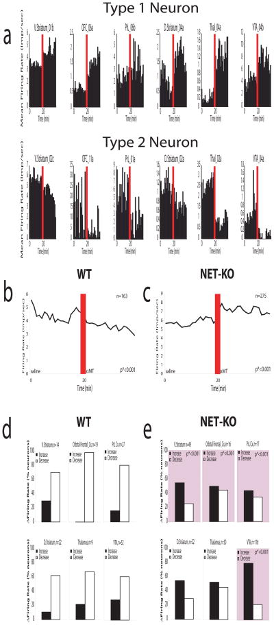
Figure 6. Norepinephrine depletion alters single unit figure rates across cortico-striatal circuits.
a) We found neurons in V. Striatum, D. Striatum, orbital frontal cortex (OFC), thalamus, and VTA that increased their firing rates following norepinephrine depletion (Type 1 Neurons), and neurons in each of these areas that decreased their firing rates following norepinephrine depletion (Type 2 Neurons). b) Treatment with αMT reduced the population firing rate observed in WT mice (n = 163; p*<0.001). c) Treatment with αMT significantly increased the population firing rate observed in NET-KO mice (n= 275; p*<0.001). d) WT mice displayed more Type 2 neurons than Type 1 neurons within each brain area investigated. e) NET-KO mice displayed a significantly decreased proportion of Type 2 neurons within V. Striatum, orbital frontal cortex, and VTA compared to WT mice (p*<0.001 for comparisons between genotype; see squares with pink highlights).

