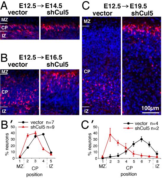
Figure 4. Cul5 regulates positioning of early neurons.
2 μg of pCAG-ChFP+Mfe or pCAG-ChFP-shCul5 DNA were microinjected in wild-type embryos at E12.5 and the position of ChFP-positive neurons inside the cortical plate analyzed at E14.5 (A), E16.5 (B) or E19.5 (C). Nuclei were stained with DAPI (blue). MZ, marginal zone; CP, cortical plate; IZ, intermediate zone. All images at same scale, bar for A-C, 100 μm.
(B’, C’) Neuron positions at E16.5 (B’) or E19.5 (C’). Mean ± SEM. Bin sizes are ~ 50 μm. Note that control neurons remain 50-100 μm from the bottom of the CP between E16.5 and E19.5, while shCul5 neurons keep pace with the top of the CP as the CP expands.

