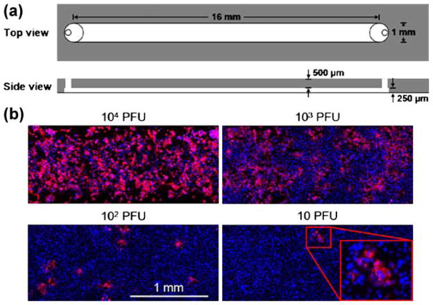
Fig. 2.
Virus infections in microchannels spread locally in the absence of flow. (a) Schematic of microfluidic channel for culture of cells and virus in the absence of fluid flow. (b) Effects of added virus level on infection patterns. Virus levels spanned from 104 plaque-forming units (PFU) per channel to 10 PFU per channel. Viral gene expression was visible by immuno-staining the viral glycoprotein (red) and cells were labeled by nuclear stain (blue). The magnified window at 10 PFU/channel highlighted a localized region of viral gene expression, initiated by the infection of one cell by a single viral particle that spread across multiple cells

