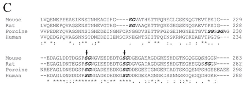
Figure 3C.

Alignment of amino acid sequences for the GAG chain attachment region of DSPP from the mouse (MacDougall et al., 1997), rat (Ritchie et al., 2001), porcine (Yamakoshi et al., 2003), and human (Gu et al., 2000). We selected a portion corresponding to mouse DSPP residues 176-283 (around the 2 GAG-linking sites) for this alignment. The names of the species are written in the left column. The amino acids are numbered in the right column. The SG dipeptides are in bold and italics. The 2 confirmed GAG attachment sites, separated by 11 amino acid residues, are marked with 2 vertical arrows. “*” indicates that the residues in that column are identical for all sequences in the alignment. “:” indicates conserved substitutions. “.” indicates semi-conserved substitutions. “-” indicates that a deletion is observed compared with the other species. Blank spaces denote the absence of any substitution, indicating that these regions are not conserved. Note that the 2 GAG-linking SG dipeptides are completely conserved among these species. There is also a high level of conservation for the 11 amino acids separating the 2 GAG-linking Ser residues as well as for the region immediately N-terminal to the first GAG-linking site, Ser242.
