
Figure 6. NQO1 residing in the mitochondrial IMS drives IB-DNQ redox cycling, promoting mitochondrial oxidative damage, Cyto c release, and activation of Casp3-mediated mitochondrial apoptotic pathway.
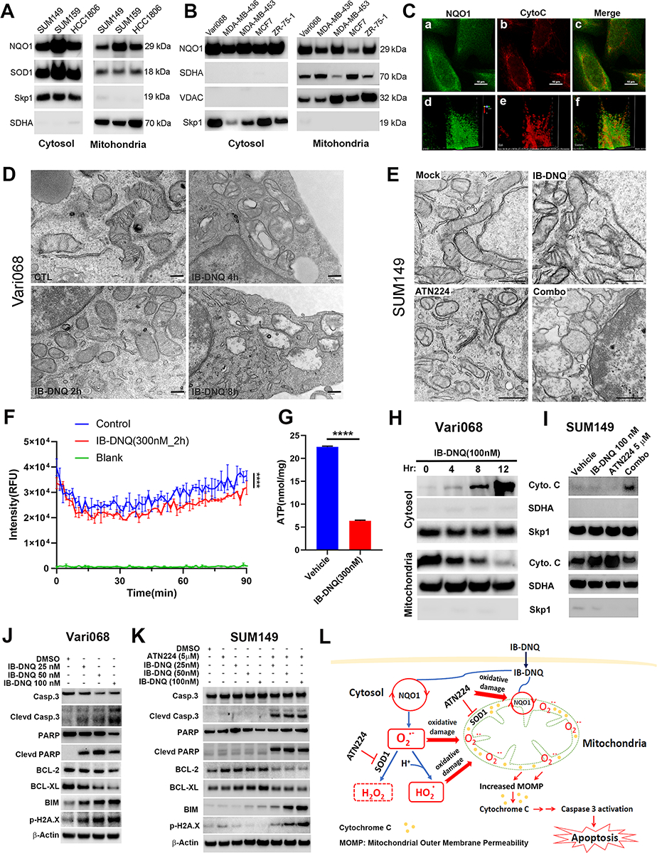
A-B, NQO1med and NQO1lo (A) and NQO1hi (B) TNBC cells were examined for NQO1, SOD1, SDHA and Skp1 expression in the mitochondrial and cytosol fraction. C, SUM159 cells labeled with specific antibodies against NQO1 and Cyto c were examined by confocal microscopy. Scale bar: 120 μm. D-E, Vari08 and SUM149 cells treated with 100 nM of IB-DNQ for 0–8h (D) or with vehicle, IB-DNQ (100 nM), ATN224 (5 μM), IB-DNQ+ATN224 for 16h (E) were processed and examined by TEM. Bars: 200 (D) and 500 (E) nm. F, SUM159 cells were treated with vehicle or IB-DNQ for 2h and measured for oxygen consumption based on fluorescent intensity (RFU) of an oxygen-bleaching fluorescent dye in the media covered by mineral oil at 1.5 min intervals for 90 min. ****: p<0.0001 (n=3, two-way ANOVA). G, ATP levels in the lysates of SUM159 cells treated with vehicle or IB-DNQ for 2h were measured and normalized by protein concentrations. ****: p<0.0001 with unpaired student’s t-test (n=3). H-I, Vari068 and SUM149 cells treated with IB-DNQ (100 nM) for 0–12h (H) or IB-DNQ and ATN224 alone or in combination for 16h (I) were used to isolate mitochondrial and cytosol fractions and examine Cyto c, SDHA and Skp1 expression. J-K, Lysates of Vari068 (J) and SUM149 (K) cells treated with IB-DNQ alone or IB-DNQ plus ATN224 for 20h were subjected to immunoblotting to examine the expression of apoptosis-related proteins. L, A model illustrating the mechanisms of action for IB-DNQ mediated pro-oxidant therapy targeting TNBC cells including CSCs.