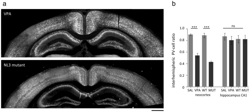
Fig 3. Interhemispheric asymmetry of PV-cell deficit in VPA and NL-3 mutant mice.
a Representative photomicrographs of PV immunohistochemistry in coronal sections of valproic acid treated (VPA; top) and Neuroligin3 mutant (NL3 MUT; bottom) mice. Note the difference in PV-cells in the two hemispheres. Scale bar: 500 μm
b Interhemispheric PV ratios calculated by dividing PV-cell numbers in the two hemispheres at the same anterior-posterior level (ratio= PV low density/PV high density hemisphere). One-way ANOVA test: *** p<0.0001; Bonferroni multiple comparison test, p<0.001; ns=not significant.

