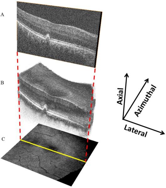
Figure 1.
SD OCT volume scan with SVP representation. B scans (A), taken sequentially at a fixed azimuthal interval (66μm) across the macula, form a volume scan (B). The three-dimensional appearance of drusen becomes apparent with volume scanning. The volume scan can be collapsed axially, with averaging of pixel intensity, to form the en face SVP retinal image (C).

