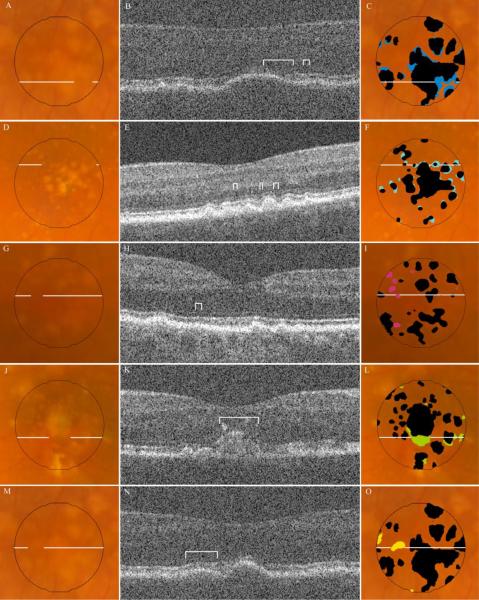
Figure 9.
Types of disagreement in drusen identification by SD OCT and CFP. Fundus photos for representative examples of disagreement are displayed in the column of images on the left, each with a line indicating location of the corresponding B-scan. The SD OCT B-scan for each sample is displayed in the middle column, with brackets identifying the region of disagreement. The column on the right displays the same fundus photo with either the SD OCT map (F, I) or composite CFP drusen map (C, L, O) in black, superimposed with color markings representing all areas of the specified disagreement type (see Table 3): A, B, C, are Type IA, undermarking of drusen borders by CFP; D, E, F are Type IB, undermarking of drusen borders by SD OCT; G, H, I, are Type II, hypopigmentation with appearance of drusen without a corresponding OCT finding; J, K, L, are Type III, pigmentary migration with obscuration of underlying drusen; and M, N, O, are Type IV, OCT deflection without corresponding CFP pigmentary change. (Not pictured: Type IC, nonspecific disagreement at drusen borders.)

