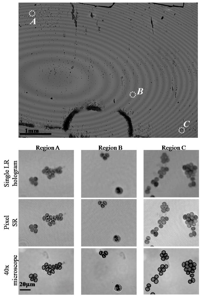
Fig. 5.
Wide-field (FOV~24 mm2) high-resolution imaging of a whole blood smear sample using Pixel SR. A comparison among the image recovered using a single LR hologram (NA<0.2), the image recovered using Pixel SR (NA~0.5), and a 40X microscope image (NA=0.65) is provided for three regions of interest at different positions within the imaging FOV. Regions (A) and (C) show red blood cell clusters that are difficult to resolve using a single LR hologram, which are now clearly resolved using Pixel SR. In region (B) the sub-cellular features of a white blood cell are also resolved.

