
Figure 1.
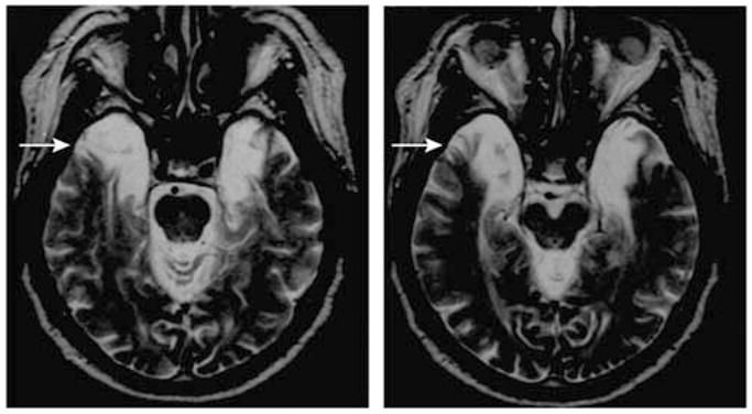
Two sections from a transverse MRI of HM’s brain illustration location of surgical lesion, which involved the excision of the rostral portion of bilateral medial temporal lobes (arrows; areas of bright signal indicate cerebrospinal fluid). Figure courtesy of Professor Suzanne Corkin.