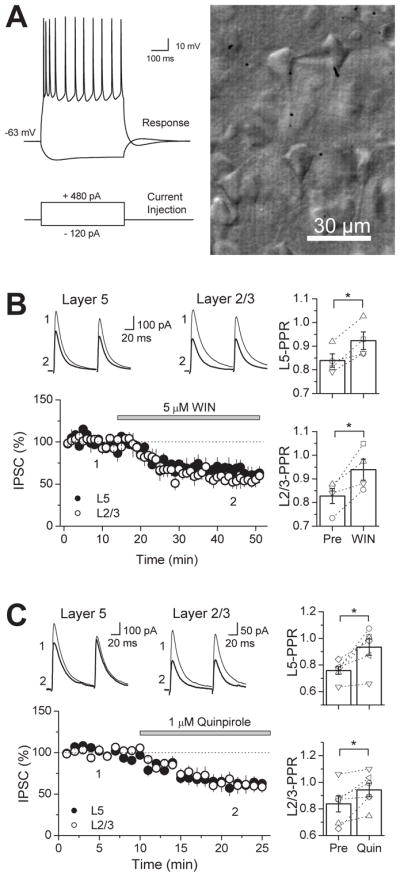
Figure 2. Pharmacological activation of CB1Rs or D2Rs suppresses GABAergic responses evoked by stimulation in layers (L) 2/3 and 5 in the mouse PFC.

(A) Left, representative response of whole-cell patched L5 pyramidal cell in current clamp (above) while depolarizing or hyperpolarizing current injections were delivered (below) to show that targeting cells in L5 by their shape is a reliable way of patching pyramidal cells. Note the initial spike-adaption, followed by a slow regular spiking behavior. Right, photograph of image under IR-DIC microscopy of a patched cell in L5 of the PFC exhibiting a pyramidal cell-like morphology. (B) Time course of WIN 55,212-2 suppression on both proximal (black circles) and distal (white circles) GABAergic inputs in the mouse PFC (n=4 cells). Top, representative average IPSC traces from a single experiment, obtained at the time points in the time-course graph. Right, bar graph of the average PPR before and after WIN application. Open symbols represent the PPR of individual experiments. (c) Time course of quinpirole suppression on both proximal (black circles) and distal (white circles) GABAergic inputs in the mouse PFC (n=6 cells). Top, representative average IPSC traces from an individual experiment, obtained at the time points indicated. Right, bar graph of the average PPR before and after quinpirole application. Open symbols represent the PPR of individual experiments. * p<0.05.
