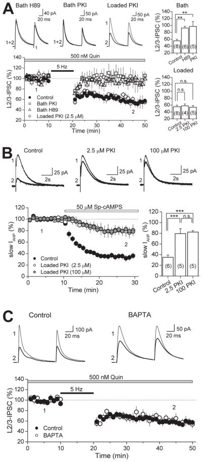
Figure 6. Neither postsynaptic PKA nor intracellular calcium rise is required for I-LTD.

(A) Time course of I-LTD in interleaved controls (black circles), bath applied H89 (10 μM; white triangles), bath applied PKI 14-22 peptide (2.5 μM; white squares) and in cells loaded with 2.5μM PKI 6-22 peptide (white circles). Inhibiting PKA activity in the slice blocks I-LTD, but inclusion of PKI (at either 2.5 μM or 100 μM) in the postsynaptic cell does not, as depicted in the summary bar plots on the top and bottom right. Top, representative average IPSC traces from single H89 and PKI experiments obtained from time points indicated. (B) Verification that intracellular loading of the PKI 6-22 peptide effectively inhibits postsynaptic PKA activity. PKI loaded into CA1 pyramidal cells at either 2.5 μM or 100 μM significantly reduced the inhibition of slow AHP current (IAHP) induced by bath application of a specific PKA activator (Sp-cAMPS). Top, representative average AHP traces from single experiments at time points indicated. Bottom right, summary bar plot depicting the effect of loading PKI on slow IAHP inhibition. (C) Time course of I-LTD in control (black circles) and in cells loaded with 20 mM BAPTA (white circles). Chelating postsynaptic calcium has no effect on I-LTD. Top, Representative average IPSC traces from single experiments. The number of experiments for each condition is indicated in parenthesis. ** p<0.01, *** p<0.005.
