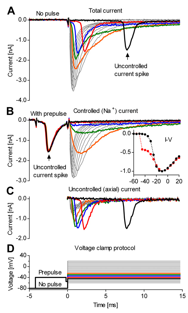
Figure 1. A voltage prepulse isolates the somatic Na+ current from neurons in slices.
Whole-cell voltage-clamp recording from a midline raphé neuron in a medullary slice. A, Steps above −50 mV evoke out-of-control current spikes. Thick traces denote currents obtained at command potentials from −45 (black) to −25 mV (orange). B, A brief pulse to −40 mV intentionally triggers the out-of-control spike, and isolates the somatic Na+ current. The inset shows the normalized peak current vs. voltage, with prepulse (black plots) and without (red plots). The prepulse restores the typical aspect of the I–V curve. C, Axial current, obtained by subtracting the currents in (B) from the currents in (A). D, Voltage clamp step protocol, with and without prepulse. To reduce the size of Na+ currents, the internal solution contained 50 mM Na+. Traces show TTX-sensitive current obtained by subtracting currents recorded before and after application of 1 µM TTX.

