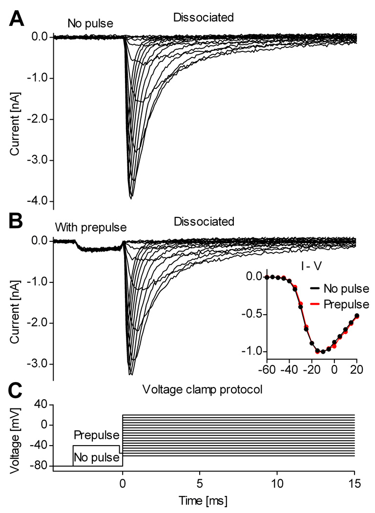
Figure 7. The voltage prepulse has minimal effects on somatic Na+ currents.
Example showing a comparison between Na+ currents recorded from a dissociated cortical pyramidal neuron, without (A) or with (B) the voltage prepulse. There is limited activation and inactivation of Nav channels during the prepulse, causing only a small reduction in the current evoked by the subsequent voltage steps (B). The inset contains the I–V curve obtained without (black plots) or with (red plots) prepulse. The traces were recorded with internal solutions containing 13 mM Na+, and were TTX-subtracted.

