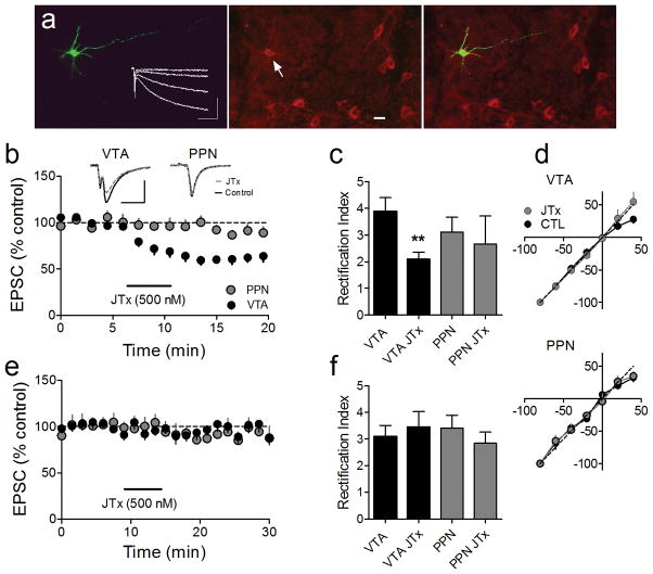
Figure 1.
Joro spider toxin (JTx) inhibition of EPSCs evoked via intra-VTA, but not PPN stimulation, indicates the absence or presence of the GluR2 AMPAR subunit, respectively, at these synapses on VTA DA neurons. (a) Photomicrograph of a biocytin-filled (left) VTA neuron that expressed the dopamine transporter (DAT, center, arrow), and a superimposition of these images (right). Also shown are membrane currents elicited by hyperpolarizing voltage steps, indicating robust Ih (left, inset). Scale bar = 20 μm. (b) Mean time course of JTx (horizontal bar) effects on EPSCs evoked via intra-VTA or PPN stimulation in the same VTA DA neurons (n = 14). Inset: representative EPSCs during control and JTx application (n = 3–5 EPSCs per average, scale bars = 10 ms, 100 pA). Data shown in b represent only neurons in which intra-VTA evoked EPSCs were inhibited by JTx (16/25, 64%). (e) Absence of JTx effect on intra-VTA or PPN-evoked EPSCs in a subpopulation of VTA DA neurons (9/25, 36%), (c). Rectification index (RI) of EPSCs evoked via intra-VTA or PPN stimulation, before and during JTx application. There was a significant decrease in RI of the intra-VTA (paired t-test, p < 0.01), but not the PPN-evoked, EPSCs in a subset of cells, whereas there was no change in the RI measured at either pathway in the remaining neurons (f, ANOVA, ns). (d) IV curves measured before and after JTx application from the same cells as in b and c.

