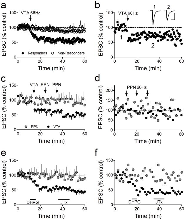
Figure 2.
Selective LTD expression at intra-VTA activated glutamate synapses. (a) Mean time course showing the effects of 66Hz stimulation of the anterior VTA on AMPAR EPSCs in all VTA DA neurons. Intra-VTA-evoked LTD was observed in 11 of 25 cells (44%, Responders). (b) Time course of LTD in a representative VTA DA neuron. Inset: mean EPSCs before and after 66Hz stimulation of the VTA (scale bars = 10 ms, 200 pA). (c) Absence of 66Hz-LTD (stimulation applied to the respective pathways at arrows) in the PPN-activated pathway, in the same VTA DA neurons demonstrating intra-VTA-initiated LTD. 66Hz stimulation was applied through the PPN electrode only when LTD was observed first in the intra-VTA pathway. LTD was never observed in the PPN pathway following 66Hz stimulation of the VTA, nor following multiple bouts of 66Hz stimulation to the PPN (arrows, and d). (d) Representative neuron demonstrating no effect of repeated 66Hz stimulation of the PPN on either PPN-or intra-VTA-evoked EPSCs. (e) Mean time course of the effects of brief DHPG application (horizontal bar) on intra-VTA and PPN-evoked EPSCs in the same VTA DA neurons. JTx (500 nM), applied after DHPG washout, had no effect on either PPN or intra-VTA-evoked EPSCs, confirming the presence of GluR2-containing AMPARs at intra-VTA-activated synapses after LTD (compare with Fig. 1b). (f) Representative example of a cell in which DHPG caused robust LTD of the intra-VTA pathway, but had no effect on the PPN stimulated pathway, and the absence of a post-LTD JTx effect.

