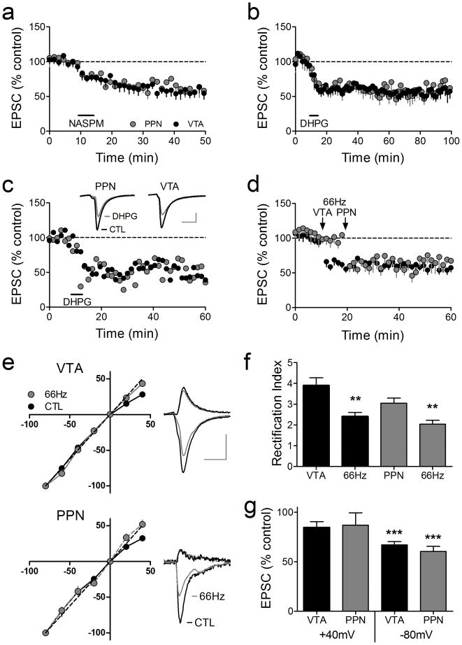
Figure 3.
A single cocaine injection changes AMPAR subunit composition, leading to reliable LTD at both intra-VTA and PPN-activated synapses in the same VTA DA neurons. (a) Mean time course of the effect of the JTx analog NASPM (horizontal bar) on EPSCs evoked via both pathways. PPN and the intra-VTA activated glutamate synapses were inhibited by NASPM in all neurons, indicating a loss of GluR2-AMPARs following cocaine exposure. (b) Mean time course of DHPG-induced LTD of EPSCs evoked via both pathways (n = 6). LTD was observed at intra-VTA and PPN activated synapses in 100% of the cells 24 hr after the cocaine injection. (c) Representative time course and EPSCs from a single DA neuron showing DHPG-LTD in both pathways (scale bars = 5 ms, 100 pA for VTA-evoked, and 5 ms, 25 pA for PPN-evoked EPSCs). (d) Mean time course showing the effect of 66Hz stimulation applied to the intra-VTA pathway, followed by PPN stimulation 10 min later (n = 8). (e) Inward rectification of EPSCs in both pathways 24 hrs following a single cocaine exposure (CTL), and subsequent loss of rectification following 66Hz stimulation in both pathways. EPSCs were normalized to those recorded at −80 mV. Example wave forms are shown at +40 mV and −80 mV before and after 66Hz stimulation in each pathway (scale bars = 5 ms, 200 pA, VTA pathway; 5 ms, 50 pA, PPN pathway). (f) Mean RI before and after 66Hz stimulation of the intra-VTA or PPN-activated inputs to the same VTA DA neurons. (g) Mean EPSC amplitude changes in both pathways at +40 mV and −80 mV after 66Hz stimulation, normalized to pre-66Hz EPSC amplitudes (−80 mV, ANOVA, p < 0.0001).

