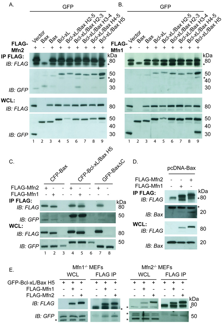
Figure 4. A chimera containing helix 5 of Bax replacing helix 5 of Bcl-xL shows enhanced binding to Mfn1 and Mfn2 relative to WT Bax and Bcl-xL.
(A–B) HeLa cells transiently expressing FLAG-Mfn2 (A) or FLAG-Mfn1 (B) and GFP vector, GFP-Bax, GFP-Bcl-xL or various GFP-Bcl-xL/Bax chimeric proteins were treated with 10µM QVD during the transfection were immunoprecipitated with FLAG conjugated beads. (C) HeLa cells transiently expressing FLAG-Mfn1 or FLAG-Mfn2 and CFP-Bax, CFP-Bcl-xL/Bax H5 or GFP-BaxΔC were immuonoprecipitated with FLAG beads. (D) HeLa cells transiently expressing FLAG-Mfn1 or FLAG-Mfn2 and untagged pcDNA-Bax were treated with 10µM QVD and immunoprecipitated with FLAG beads. (E) Mfn1−/− MEFs or Mfn2−/− MEFs transiently expressing FLAG-Mfn2 or FLAG-Mfn1 and GFP-Bcl-xL Bax H5 were immunoprecipitated with FLAG beads. FLAG bead immunoprecipitate (IP) and whole cells lysate (WCL) samples were analyzed using FLAG and GFP antibodies (A–C and E) or FLAG and Bax antibodies (D). Asterisks represent non-specific bands. These data are representative of at least two independent experiments.

