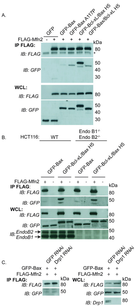
Figure 5. Co-immunoprecipitation of GFP-Bax and GFP-Bcl-xL/Bax H5 and the mitofusins.
(A) HeLa cells were transfected with FLAG-Mfn2 and GFP, GFP-Bax, GFP-Bax A117P (mutation within H5), GFP-Bcl-xL/Bax H5 or GFP-Bax/Bcl-xL H5 (the reverse H5 chimera) and immunoprecipitated with FLAG beads and blotted for FLAG and GFP. (B) WT HCT116 and EndoB1−/− EndoB2−/− HCT116 cells transfected with FLAG-Mfn2 and GFP-Bax or GFP-Bcl-xL/Bax H5 were immunoprecipitated with FLAG beads and blotted for FLAG, GFP, Endophilin B1 and Endophilin B2. (C) Drp1 RNAi or control GFP RNAi HeLa cells (31) were transfected with FLAG-Mfn2 and GFP-Bax and immunoprecipated and blotted as in (A). Drp1 protein level after RNAi treatment was analyzed using anti-Drp1 antibody. Asterisks represent non-specific bands. These data are representative of at least two independent experiments.

