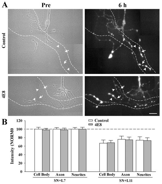
Figure 5.
Reducing ApCAM from the surface of the sensory neurons does not affect target induced regulation of sensorin expression. Cultures were processed as described above, except at 6 h the co-cultures were fixed and processed for sensorin immunochemistry. A. Phase contrast images (left) of axon growth from the stump of sensory neurons treated overnight with control IgG (n = 7 for SN + L7 or SN + L11) or 4E8 (n = 7 for SN + L7 or SN + L11). The dashed outline represents the position of the motor neuron L7 (axon hillock and major processes) that was plated with each sensory neuron after taking the images (Only SN + L7 cultures are shown here). Treatment with 4E8 did not affect sensorin expression in the regenerated neurites, especially the new branches and varicosities. On the right are epifluorescent images of sensorin immunostaining from the same sensory neuron at 6 h of interacting with the motor neuron. In both images arrowheads point to the location of some of the varicosities and arrows point to the location of some of the new branches that had grown over the 6 h of interaction. The bar equals 40 μm. B. Reducing ApCAM from the surface of the sensory neurons did not block target-induced regulation of sensorin expression throughout the sensory neurons. The height of each bar is MEAN ± SEM of the staining intensity in the 4E8-treated cultures after normalization with the staining intensity for each compartment of sensory neurons treated with controls and interacting with L7 after 6 h. Treatment with 4E8 failed to block the strong staining in sensory neurons contacting L7, and failed to block the reduction in staining in each compartment for sensory neurons contacting L11.

