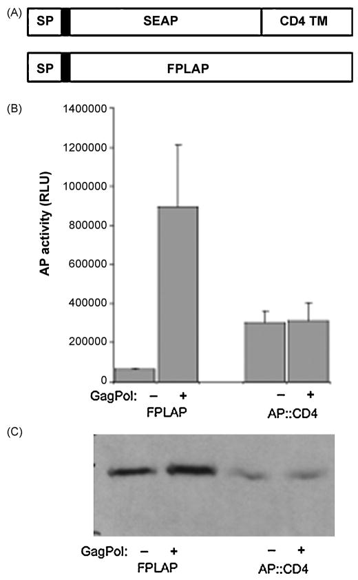
Figure 2. Incorporation of alkaline phosphatase activity in Gag-derived virus-like particles.
A, Schematic representation of AP::CD4 and FPLAP. The Flag-tag is indicated by a black box following the signal peptide (SP). TM, transmembrane domain. B, 293ET cells were transiently co-transfected with MLV Gag-Pol and FPLAP or AP::CD4. Supernatants were harvested after 3 days and analyzed for alkaline phosphatase activity and compared to cells that were transfected with FPLAP constructs only. Results from three independent infections + standard deviation (SD) are shown. RLU, relative light units. C, Cell lysates from A were analyzed by immuno-blotting of FPLAP and AP::CD4 in the presence or absence of Gag-Pol using anti-Flag M2 antibody.

