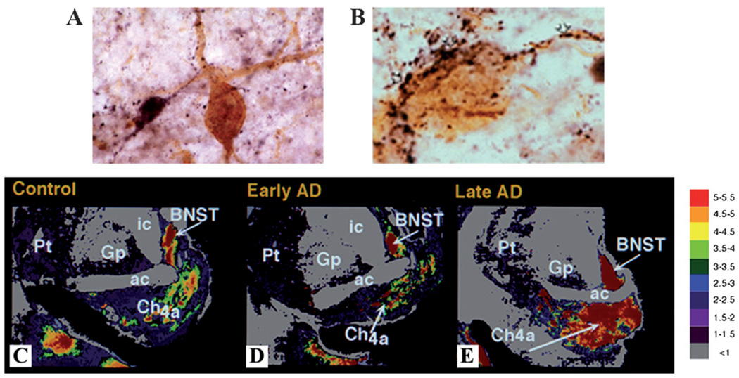
Figure 1.
GAL plasticity in the basal forebrain nucleus basalis in AD. (A) Photomicrograph shows a magnocellular cholinergic NB neuron immunostained with the CBF neuronal marker p75NTR (brown reaction product) and innervated by GAL-ir fibers (dark blue reaction product) in aged control brain. Note the GAL-ir parvicellular neuron contacting the CBF neuron. (B) Photomicrograph shows a striking hyperinnervation of GAL fibers impinging upon a cholinergic NB neuron in AD. (C–E) Pseudocolor density maps showing the regional distribution of [125I]hGAL binding sites in the aged control NB (C) as compared to early stage (D) and late stage (E) AD. Note the increase in labeling in the anterior subfield of the NB in late AD. Gray to red on the color scale indicates increasing GAL binding levels. AD, Alzheimer’s disease; NB, nucleus basalis; CBF, cholinergic basal forebrain. ac, anterior comminsure; BSNT, bed nucleus of stria terminalis; Ch4a, NB anterior subfield; GP, globus pallidus; ic, internal capsule; PT, putamen.

