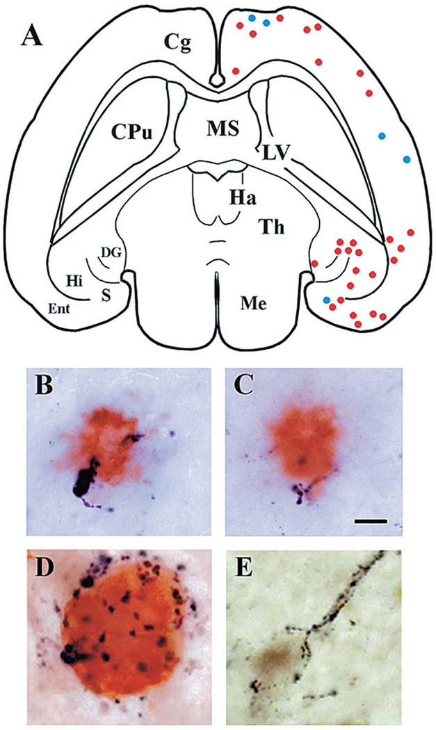
Figure 2.
Association of GAL with AD-related lesions. (A) Schematic drawing of a horizontal brain section from a 3-month-old APPswe/PS1ΔE9 transgenic mouse showing the distribution of Aβ-ir plaques with adjacent GAL-ir dystrophic neurites (blue dots) or without dystrophic neurites (red dots). Cg, cingulate cortex; CPu, caudate-putamen; DG, dentate gyrus; Ent, entorhinal cortex; Ha, habenula; Hi, hippocampus; LV, lateral ventricle; Me, mesencephalon; S, subiculum; Th, thalamus. (B, C) Photomicrographs show co-localization of GAL fibers (black) and Aβ (orange) in amyloid plaques located in the cortex (B) and hippocampus (C) of a 3-month-old APPswe/PS1ΔE9 mouse. Scale bar, 10 µm. (D) GAL hyperinnervation (black) of a cholinergic NB neuron dual-stained for Tau C3 (orange), a tau epitope that appears early in the evolution of NFTs. (E) GAL hyperinnervation (black) upon a cholinergic NB neuron immunonegative for MN423, a late-stage tau event in NFT formation. There was no evidence for GAL hyperinnervation of MN423-immunopositive CBF neurons.

