
Figure 4. Sensitivity of EA.hy926 endothelial cells to vinblastine and pegylated IFN-α.
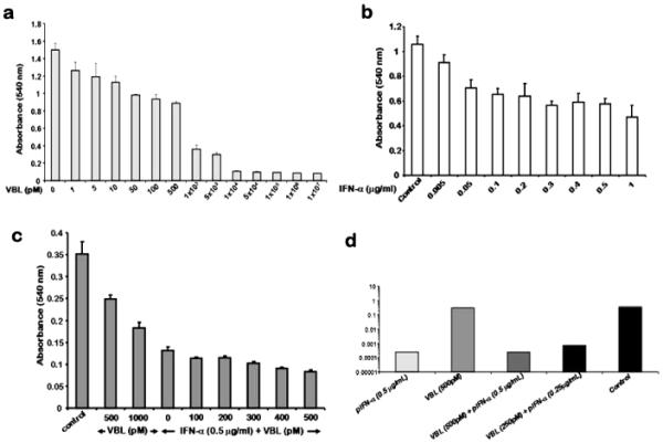
3000 cells/well in a 96-well plate were treated with indicated concentrations of vinblastine (a) or pegylated IFN-a2b (b) for 72 h and subjected to MTT colorimetric assay. (c) Cells were treated with the pegylated IFN-a2b (0.5 mg/ml) in combination with the indicated concentrations of vinblastine (0 - 500 pM) for 72 h. Cell viability was estimated by MTT assay. The above three figures were representative of three independent experiments the results of each are the means ± standard deviation (n = 6), (d) 2000 cells/well in a 6-well plate were treated with the indicated concentration of drugs for 24 h. The cells were rinsed, fresh medium was added and after 2 weeks, colonies were stained with crystal violet, counted and surviving fraction calculated.