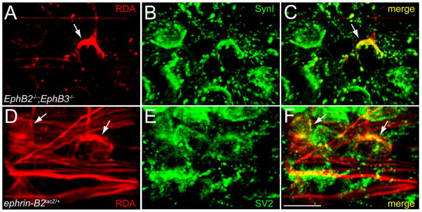
Figure 5. Aberrant ipsilateral calyces express presynaptic markers.
A. In this mutant ipsilateral MNTB (P9 EphB2−/−;EphB3−/−), RDA labeling reveals an aberrant calyx (arrow). B. Expression of synapsin I in the same section as in A. C. Merge of panels A and B indicates that the RDA-labeled aberrant calyx is also positive for synapsin I (yellow). D. In a different mutant ipsilateral MNTB (P26 ephrin-B2lacZ/+) RDA labeling reveals axons (red) and two aberrant calyces (arrows). E. SV2 immunofluorescence in the same section as in D. F. Merge of panels D and E indicates that the RDA-labeled calyces (arrows) are also positive for SV2 (yellow). Scale bar in F also applies to A–E, and indicates 20 μm.

