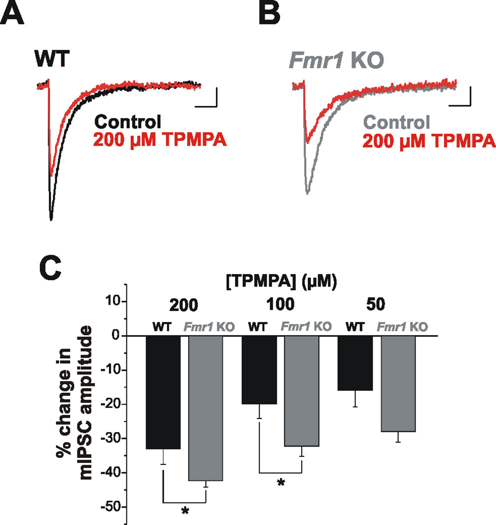
Figure 4.
Synaptic GABA release is reduced in Fmr1 KOs. (A–C) mIPSC amplitude is reduced to a greater extent by TPMPA in Fmr1 KO than in WT mice. Averaged mIPSCs from individual cells from WT (A) and Fmr1 KO (B) under control conditions and with TPMPA (200 µM) (red traces) illustrate the reduction in amplitude. (C) Averaged group data indicating that TPMPA had a significantly greater effect on mIPSCs in Fmr1 KOs at doses of 200 µM and 100 µm. This trend was also observed using a 50 µM dose, but was not statistically significant. TPMPA did not exert a significant effect on mIPSC decay. *p < 0.05. Scale bars: 2 pA, 20 ms.

