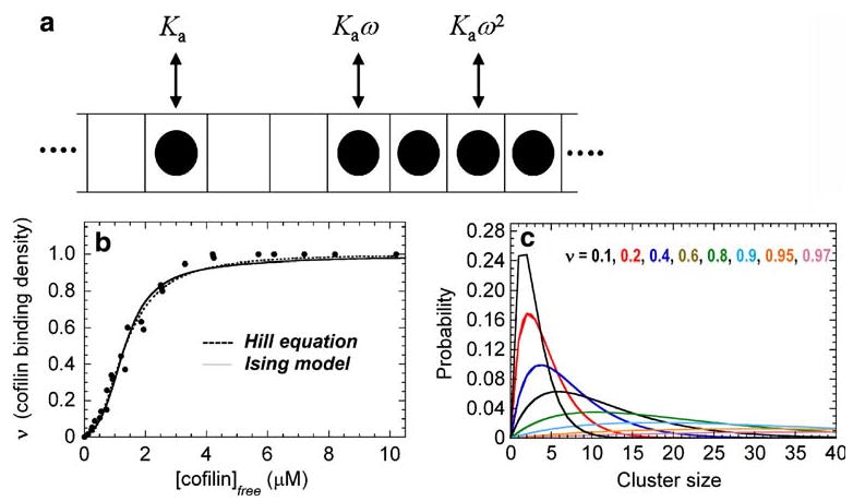
Fig. 1.

a Schematic of one-dimensional Ising model of cofilin binding to an actin filament. Individual filament subunits are depicted as squares of an infinite, one-dimensional (1-D) lattice. A subunit with bound cofilin is indicated with a filled circle. Ka is the association equilibrium binding constant, and ω is the unitless cooperativity factor. The overall binding constants are given by Ka (isolated, non-contiguous bound cofilin), Kaω (singly-contiguous bound cofilin) and Kaω2 (doubly contiguous bound cofilin). b Cooperative cofilin binding to actin filaments. The lines represent the best fit of the data for human cofilin-1 binding to rabbit muscle actin (filled circles) to the Hill equation (dotted line) or binding to a 1-D lattice with nearest neighbor interactions as depicted in a. The figure is adapted from De La Cruz (2005). c Cofilin cluster size distribution. The probability of bound cofilin being in a given cluster size. Each line represents the distribution at the color-specified binding density
