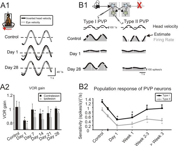
Figure 2.
Changes in simultaneously measured VOR and neuronal responses after unilateral labyrinthectomy. (A1) Example VOR responses show reduced and asymmetric gains immediately following lesion (day 1), and impressive compensation, recovering to nearly normal values by day 28. Note, head velocity traces have been inverted to facilitate comparison with the evoked eye velocities. (A2) VOR gains averaged across both animals. On day 1, responses were reduced for both ipsi and contralaterally directed rotations. Over the next 2–3 weeks, contralesional gains improved to normal values and ipsilesional gains were nearly compensatory. (B1) Examples of type I and type II PVP responses before and at different time points after contralateral labyrinthectomy. Response of both cell types decreased significantly immediately following lesion (day 1). While the sensitivity of type I neurons improved over time reaching normal values by day 28, that of type II neurons did not show significant improvement (P> 0.1). Inset shows that type I neurons receive indirect inputs from contralateral labyrinth through inhibitory type II neurons. (B2) Summary of the analysis of i) the population of 57 neurons (40 type I and 17 type II) recorded under control conditions and ii) the population of 109 neurons recorded after lesion (56 type I and 53 type II). Of the latter group, 44 were recorded on the first day (i.e., 15–28 hours) post-lesion, 32 in the period of 7–21 days post-lesion, and 33 in the 1–2 months post-lesion. * represent significant difference re. control (i.e., before lesion), P<0.05

