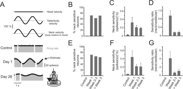
Figure 3.
The majority of PVP neurons show robust modulation to stimulation of neck proprioceptors after contralateral labyrinthectomy. (A) Examples of type I neuronal responses during stimulation of neck proprioceptors. In intact animals, neurons are not sensitive to stimulation. In contrast, the example neuron shown on day 1 after the lesion responded robustly to neck stimulation. The neuron shown on day 28 also responded to neck stimulation, but with a lower sensitivity. (B) The percentage of neck-sensitive type I neurons remained constant (60–70%) from week 1 to 8 after lesion. (C) The average of the absolute values of neck sensitivities of type I neurons decreased during compensation, but never reached control values (i.e., responses remained significant). (D) Decreases in neck sensitivity of type I neurons were temporally linked to increases in the vestibular sensitivities. Accordingly, neck sensitivities were the most robust the first week after lesion. (E) The percentage of neck sensitive type II neurons remained constant (60–70%) from week 1 to 8 after lesion (n=29). (F) The average of the absolute values of neck sensitivity in type II neurons decreased from 0.44±0.15 on week 1 post-lesion to 0.22±0.05 and 0.20±0.08 during weeks 2–3 and after week 3, respectively. (G) The ratio of neck and vestibular sensitivities shows that neck sensitivities of type II neurons were the most robust the first week after lesion.

