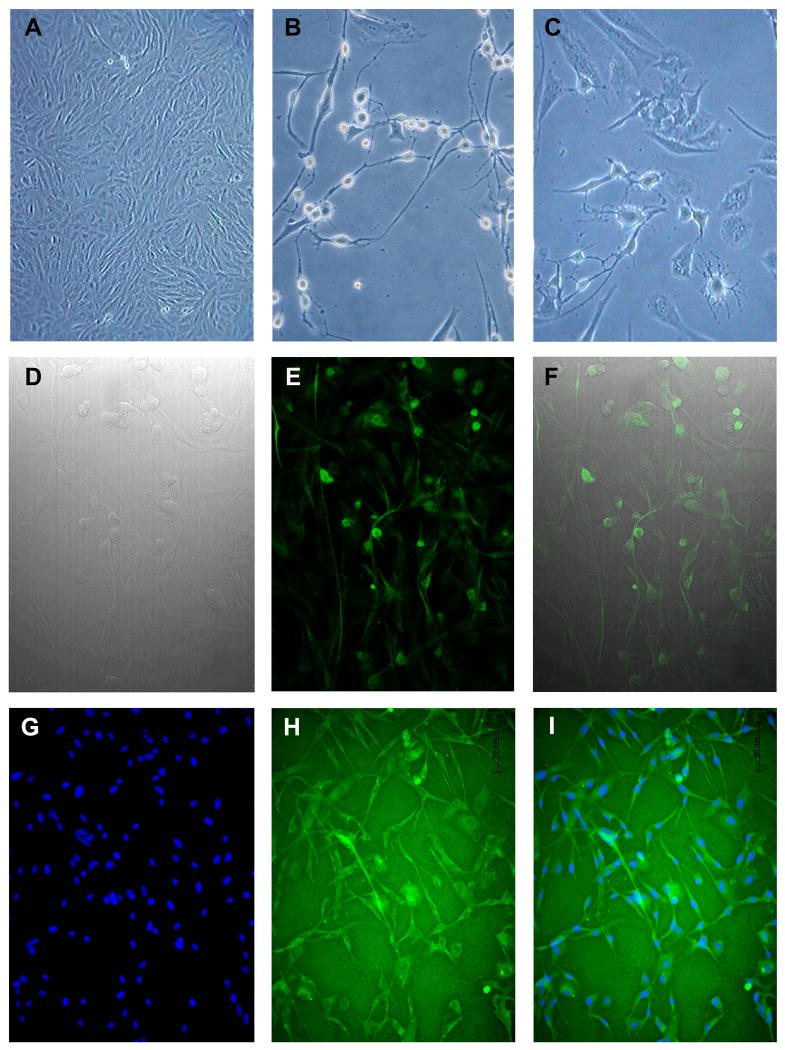
FIG 1. In vitro Neurogenic Differentiation of HEDSC.
HEDSC cultured in control media demonstrate typical stromal cell morphology (A), while cells cultured in neurogenic media demonstrated both pyramidal and dendritic cell morphology as is pictured using light microscopy (B, C). Differentiated cells visualized using: Differential Interference Contrast (DIC) (D), IF for neural stem cell marker Nestin expression (E), and a merge of both (F). Differentiated cell cultures, also express tyrosine hydroxylase (H), DAPI nuclei staining (G), and merge of both (I).

