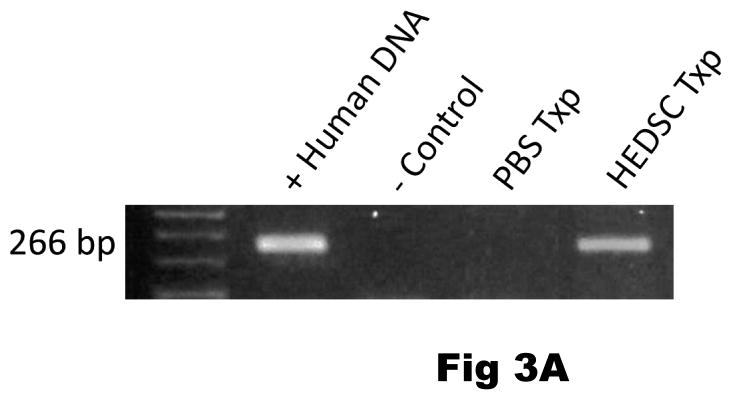
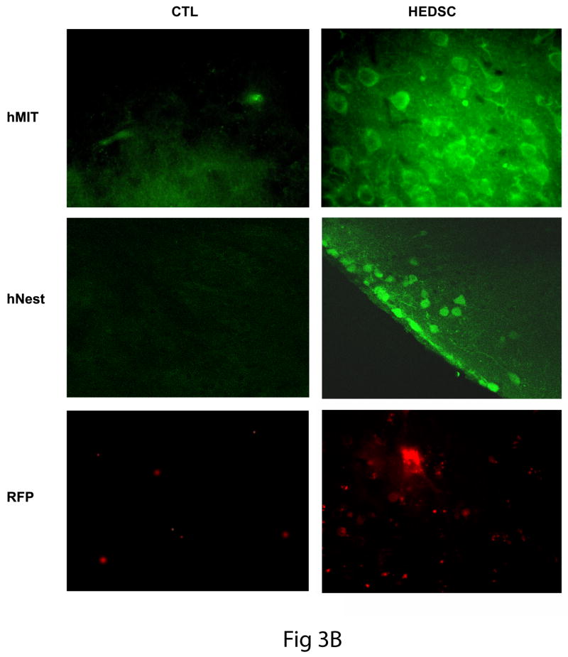
FIG 3. HEDSC Engraft, Differentiate in vivo, and Migrate to the Lesioned Site in Mice Brains.
(A) PCR detecting human DNA in mouse brain transplanted with HEDSC (n=14), but not with sham transplants (n=8) using PBS. (B) Low power view of murine brain section from sham treated (CTL) and HEDSC treated animals. An area that includes the SN is outlined. IHC using a human nestin antibody identifies cells localized to the SN in the transplanted animals (C) Human cells are visualized in mouse brains in the right column, while controls are shown on the left. In the top panel, all human cells are detected using a human mitochondrial antibody (hMit), which are seen here at the site of transplantation in the striatum. Spontaneous in vivo differentiation of transplanted HEDSC was observed, where they expressed Nestin (hNestin). Transplanted cells adapted a neurogenic phenotype morphologically, as is visualized using red fluorescent surface labelling (RFP). Human cells were observed remote from the initial transplantation site (striatum), where they migrated to the lesioned brain area (substantia nigra) which is the area pictured in the bottom right panel (RFP).


