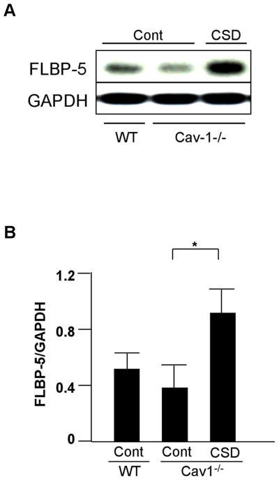
Figure 7.
IGFBP-5 uptake following restoration of Cav-1 function. A: WT or Cav-1−/− fibroblasts were treated with Cav-1 CSD peptide (CSD) or control peptide (Cont) for 1h in serum-free medium, then FLAG-tagged IGFBP-5 (FLBP-5) was added. After 15 minutes, lysates were harvested and subjected to western blot analysis. IGFBP-5 levels in lysates were detected using anti-FLAG M2 antibody. GAPDH was used as a loading control. B: Graphical analysis of IGFBP-5 levels in lysates of WT and Cav-1−/− fibroblasts following 1h incubation with Cav-1 CSD (CSD) or control peptide (Cont). Horizontal bars indicate mean values from 4 independent experiments. The paired t-test was used for statistical analysis. * P < 0.05.

