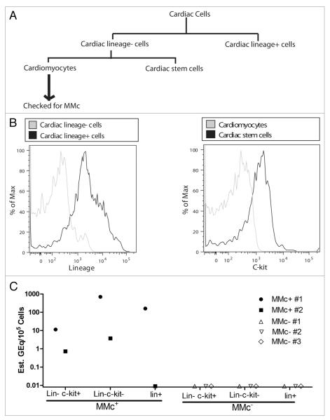
Figure 3.
MMc in cardiac stem cells is associated with MMc in cardiomyocytes. (A) Lin+ cells were sorted from cells extracted from the hearts of NIMAd-exposed offspring using a MACS sorter. C-kit+ cells (cardiac stem cells) were sorted from the lin− fraction. The lin−c-kit− cells are mostly cardiomyocytes. The cells were further sorted using a FACS sorter to obtain 100% purity. Cardiomyocytes were checked for MMc. Different cell fractions isolated from the hearts of the offspring having detectable or undetectable MMc in the cardiomyocte fractions were pooled and represented as MMc+ and MMc−, respectively. (B) Different cardiac cell populations after MACS sorting. (C) DNA was extracted from the different cardiac cell populations from MMc+ and MMc− offspring. They were checked for MMc. Each dot represents MMc level in a pooled cell fraction of 4–5 NIMAd-exposed offspring.

